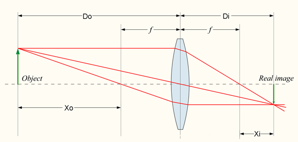
Equation 1: 1/Do + 1/Di = 1/f
Equation 2: Xo * Xi = f*f
Equation 3: M = - Di / Do = f / (f-Do)
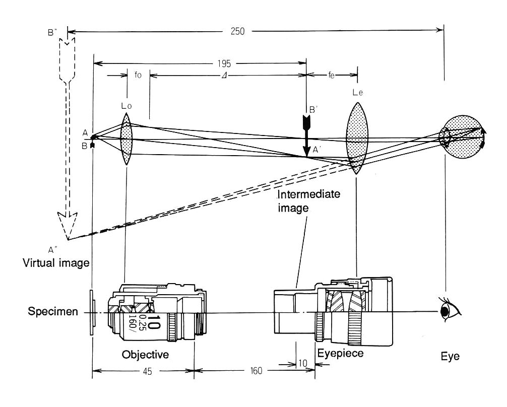
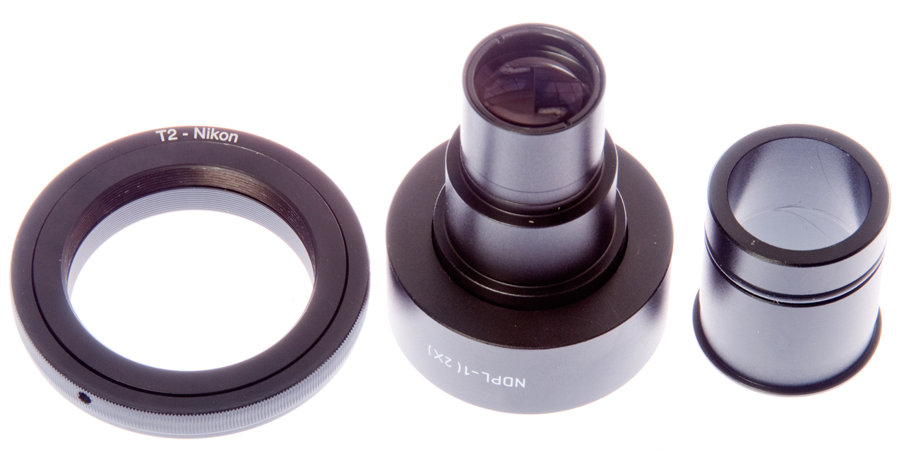
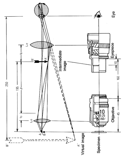
Microscope 160mm Tube Length Optics diagram (from Nikon booklet How
To Use A Microscope and Take a Photograph
Note that the parafocal distance is shown as 45mm which indicates
this is applicable to Nikon Chrome Free (CF) optics.
It also shows the real objective image 10mm below the top of the
tube, i.e. at 150mm from the objective flange.
fo: Objective Focal Length
fe: Eyepiece Focal Length
45mm Objective Parafocal distance (i.e. CF optics)
160mm tube length
195mm from subject to real image
205mm from subject to top of tube
Lo: plane of the objective
Le: plane of the eyepiece
A-B subject on top of slide and below cover glass
A'-B' Real Image from objective 10mm below top of tube
A"-B" virtual image as seen by eye (250mm from eye)
FL (objective) = 160mm / power
Example:
10x objective is 16mm focal length.
FL (eyepiece) = 250mm/(power-1)
Example:
10x eyepiece is 27.77mm focal length
Note: The 250mm shown in the diagram is
really 10 inches converted to metric with
rounding. Based on The Microscope and it's
Revelations by Carpenter 1856
|
Nikon Microscope CF Optics, 160mm Tube
Length, 45mm Parafocal Distance
|
Numerical Aperture (Wiki:
N.A.)
Camera lenses (Wiki)
are marked with f/ numbers which are calculated by dividing
the focal length by the effective lens diameter.
For example for the Nikon 300mm (focal length) lens that is
f/2.8 the lens diameter is FL/f = dia = 107mm or 4-1/4".
The lower the f/number the "faster" the lens, i.e. the better
it works in low light conditions.
Also the lower the f/number the higher resolution if the lens
design is diffraction limited (Wiki).
The idea of diffraction limited optical systems applies to
microscopes, telescopes, cameras, radio telescopes, etc. but
not to low cost optical devices.
The diffraction limited spot size in micro meters for a lens
is close to the f/# divided by 2. So for the Nikon 300mm
f/2.8 lens the spot size when at f/2.8 would be 1.4 um.
Note for astronomical telescopes if you want small spot size
you need a low f/#. As a lens is stopped down to higher
f/numbers the spot size gets larger, i.e. the resolution gets
poorer. But the depth of field gets deeper. So
when you stop down a camera in order to get a greater depth of
focus you are at the same time getting less resolution.
If you want both resolution and depth of focus then focus
stacking is the way to go.
The numerical aperture (N.A.) is a measure of the resolving
power of a lens used in air and when focused at infinity, like
an astronomical telescope or camera it is:
N.A. = D / (2 * FL) for the Nikon 300mm f/2.8 lens it
would be 107mm / (2 * 300mm) = 0.178
The N.A. is defined as n * SIN(theta)
where:
N.A. is a dimensionless number
n is the refractive index (air=1, water = 1.33, objective
immersion oil = 1.515)
theta is the half angle of the triangle formed by the point in
focus and the diameter of the objective lens. For
example if the diameter of two objectives are different and
the working distance between the lens and the subject is the
same the lens with the larger diameter will have a higher
N.A. In a similar manner if two objectives have the same
lens diameter and one of them has a smaller working distance
it will have the larger N.A.
Note this is exactly the same as a camera where for a given
focal length lens the one with the larger diameter has higher
resolution and lower f/number.
The resolution (Wiki)
of a microscope is given by:
Ernst Abbe (Wiki)
derived the math relating to microscope N.A. and invented the
condenser named after him (Wiki: Abbe
Condenser). He was the optical designer at Carl
Zeiss (Wiki: the man,
the
optical company) and developed their microscopes and
also invented the three color corrected lens (Wiki: Apochromat).
The overall magnification of a microscope is
the product of the objective power and the eye piece power.
The range of useful magnification depends on the N.A. of the
optical system and is given as between 500 and 1,000 times
the system N.A.(when viewed by human eyes).
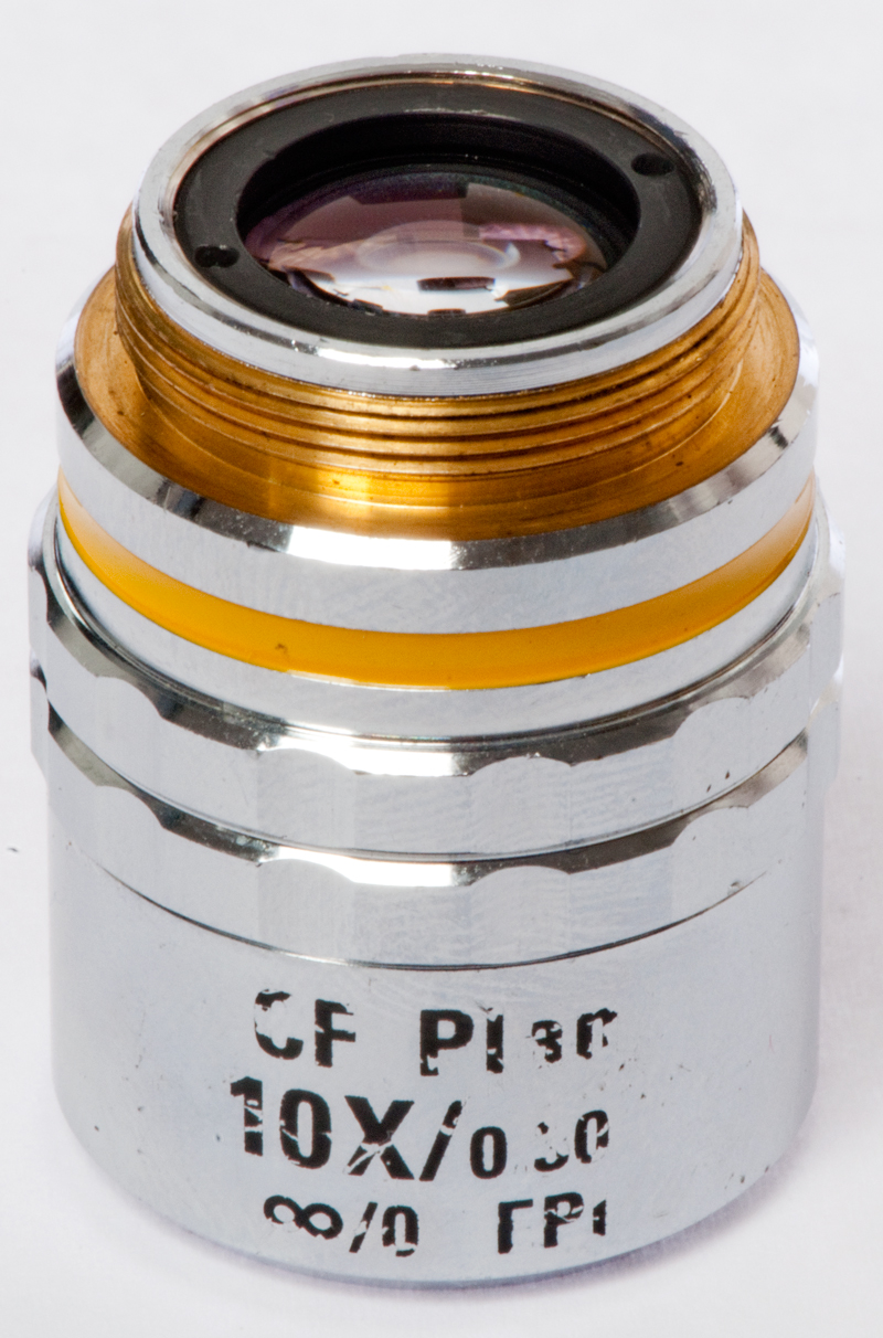
For example for the Nikon CF
Plan 10x/0.30 ∞ / 0 EPI objective the magnification could be
between 150X and 300X.
If the overall magnification is below 500 * N.A. then you
are not seeing all that you could.
If the overall magnification is above 1000 * N.A. then
you have what's called "hollow magnification", that's to say
the image is larger but there's not an increase of
detail. The above guidelines are based on the
resolution of the human eye (Wiki: 4
arc minutes)
When a camera is used there are
similar guidelines based on the number and size of the
camera's pixels.
Digital
Camera Resolution Requirements for Optical Microscopy -
This page says for the Nikon 10X / 0.30 N.A. objective the
resolution limit is 0.92 um, projected size is 9.2 um and the
required pixel size is 4.6 um.
The Nikon D300s has 5.5 x 5.5 um pixels ( 4288 x 2848) so it's
pretty close. But for the whole range of objectives the
required pixel size varies between 3.5 um (1X/0.04) to 10 um
(100X/1.40) so the D300s is a reasonable fit to all of
them.
Although a board CMOS camera that has pixels that are a 6 x 7 um
it's resolution of 728 x 488 pixels is much lower than the Nikon
D300s.
The big advantage of this method is much lower cost than using
a modern microscope which can easily cost thousands of dollars,
whereas the adapters and objectives for this method and in the
hundreds of dollars. The disadvantage of this method is it
may be difficult to use modern lighting methods that are much
easier with a modern microscope. There's also the question
of using infinity lenses because of the need for a "tube
lens". I have a couple of doublets on order to see how
hard it is to use infinity lenses in this setup.
A camera mount that may be suitable for this is the Nikon Multiphot. Photo studio
high power strobe
lighting (used for Fig 3 below) should also work.
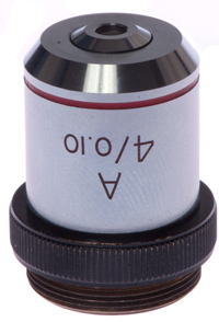
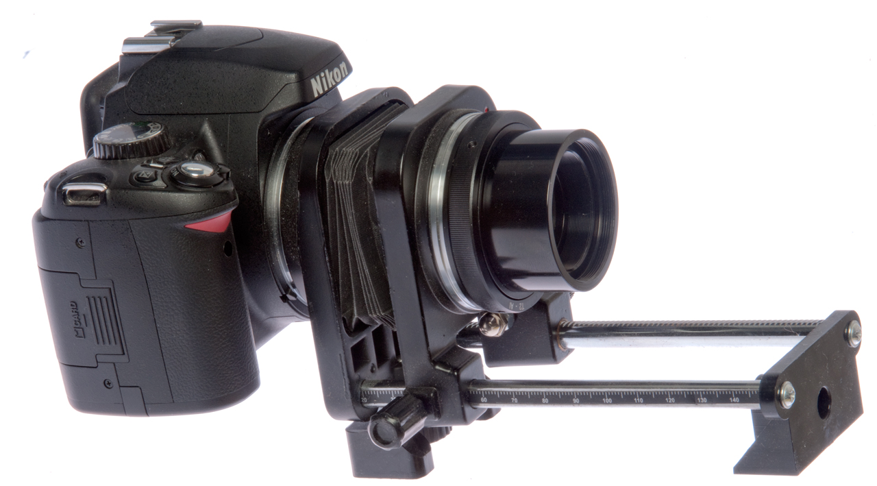
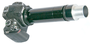
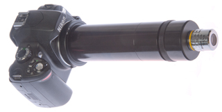
It turns out that the Nikon D60 (shown in Fig 2 below) is NOT a
good choice for this type of photography because it does NOT
have live view. You can focus by looking through the
normal TTL viewfinder, but there's no way to magnify the image
(maybe with an optional magnifying viewfinder
attachment?). But when the D300s is used and in live-view
the (+) bottom is pressed a few times you can see very fine
detail which allows focusing.
So, to use this method it's easier if you have a camera that
supports live-view.
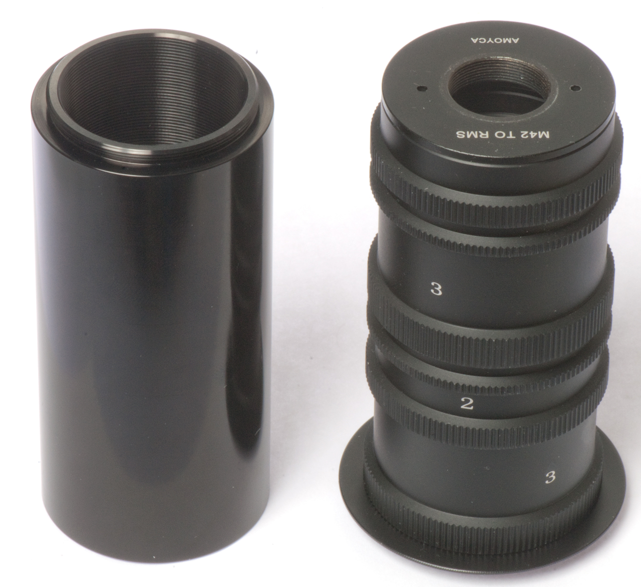
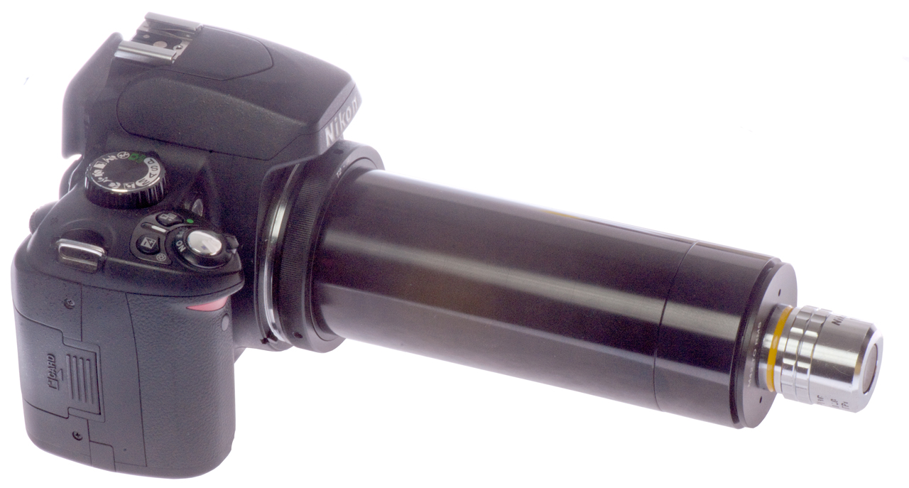
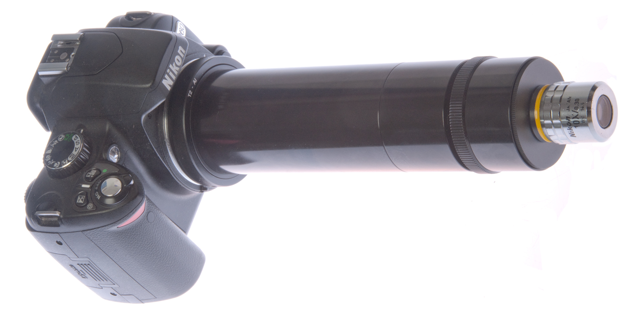
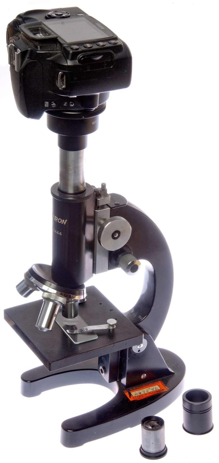
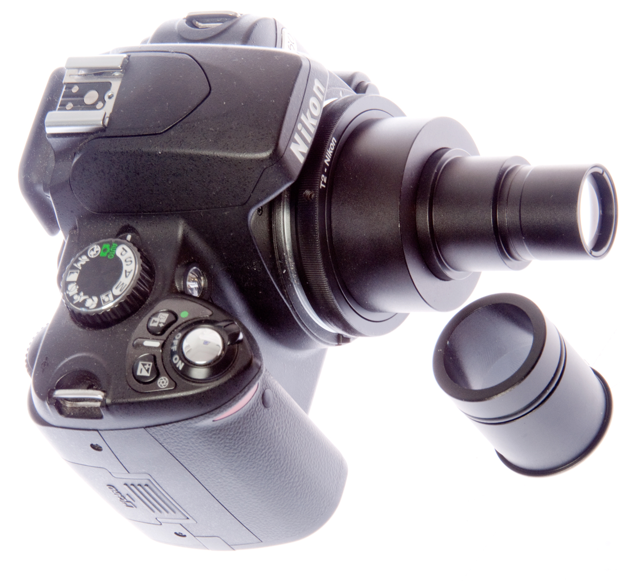
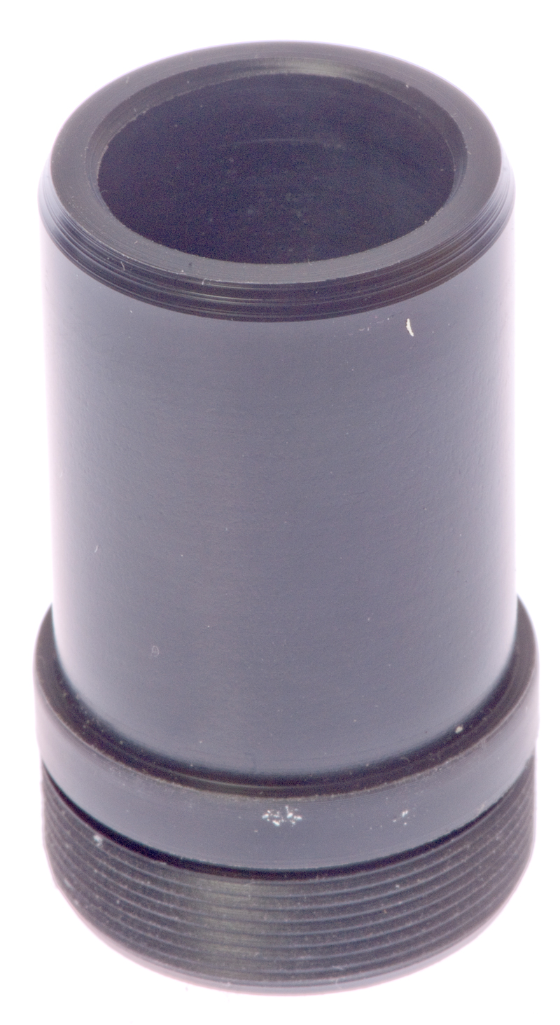
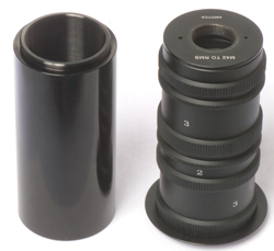
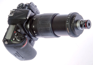
Fig 1 Adapters Nikon to Objective

|
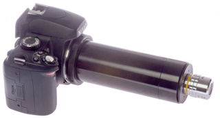
Fig 2 Nikon D60 shown with all 8 extension
tubes & Objective.
|
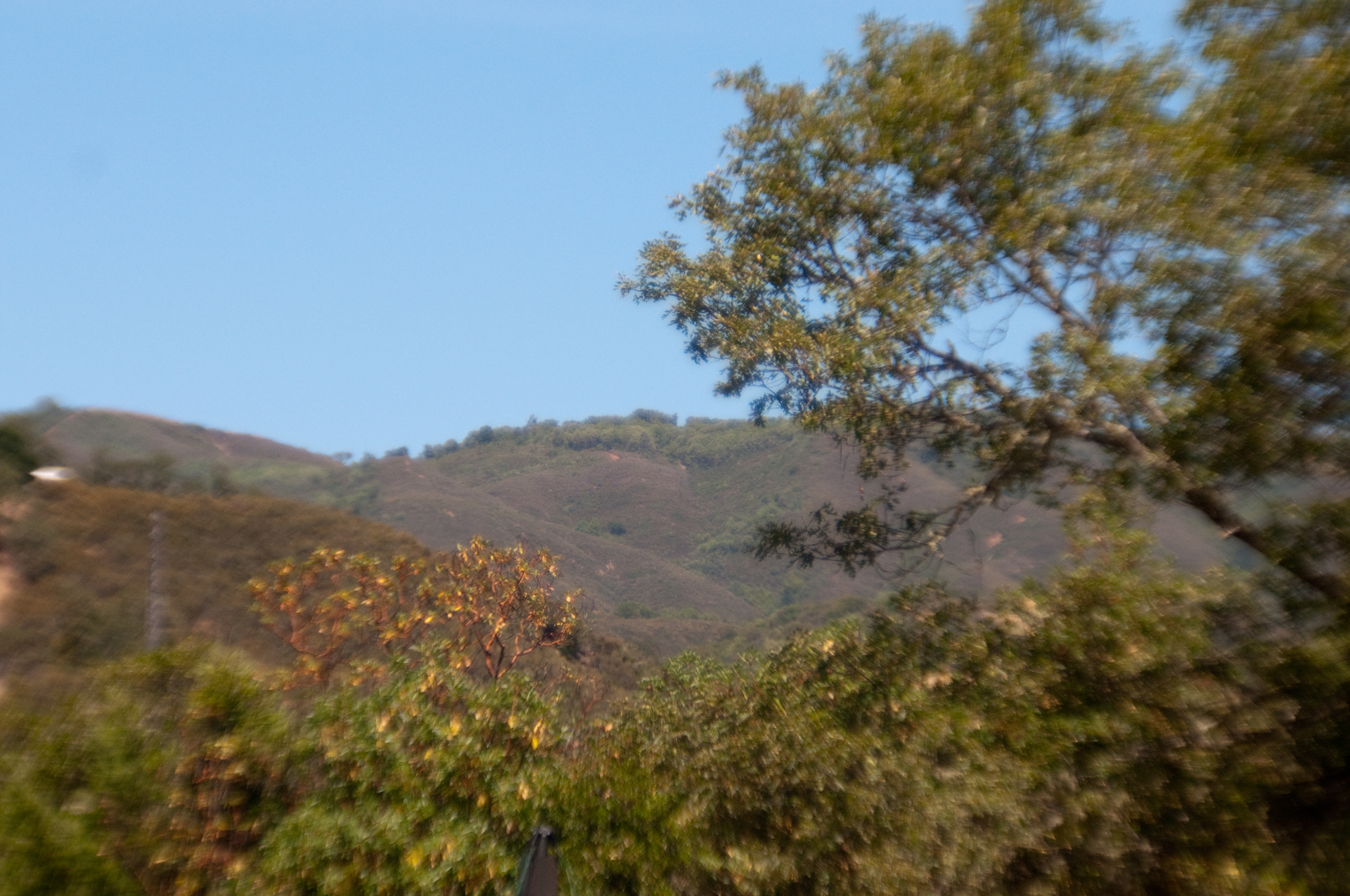
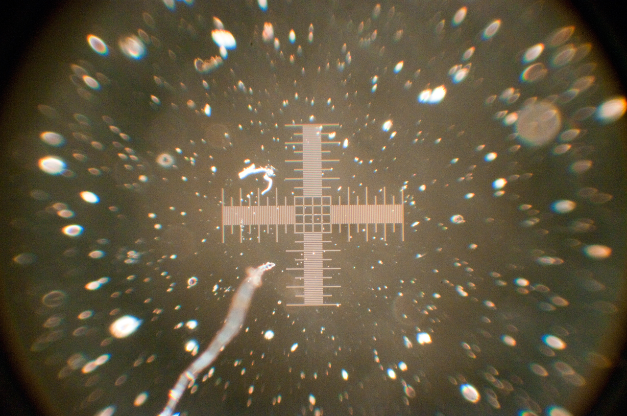
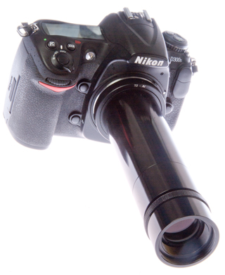
Fig 3 sample photo Nikon D300S DX handheld
all 8 extension tubes
|
Fig 4 T-mount (42mm) to 25mm Objective
adapter
|
Fig 5 T-Mount (42mm) Extension Tubes each
stack is close to100mm
The T-mount threads on the extension tubes do not match
the
Edmund Optics 100mm extension tube.
|
Fig 6 "Chipped" Nikon T-Mount adapter
Front
Back
These threads match the Edmund Optics parts.
|
The adapters are:
Nikon camera body to T-Mount (aka: 42mm) (Wiki)
Extension tubes (two sets) length includes camera to T-mount
(0.89mm) and T-mount to RMS (2.91 round to 3 mm)
Tube 1: 7mm
Tube 2: 14mm
Tube 3: 28mm
T-mount to RMS microscope
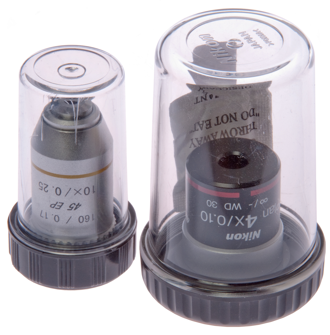
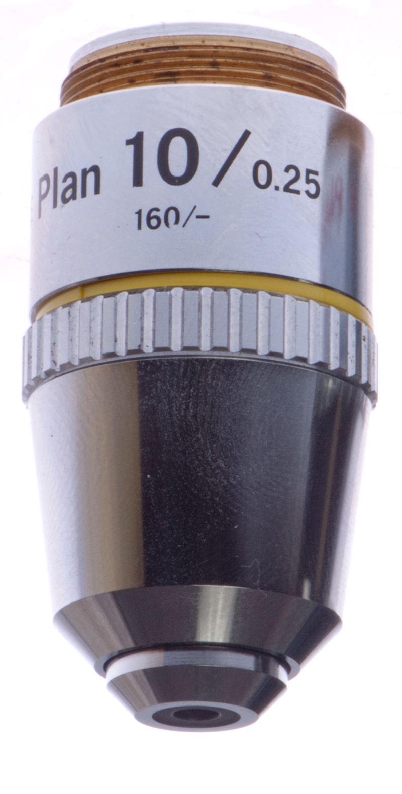
10X objective 160/0.17, 45 EP, 10X/0.25 Translation: 160mm tube
length/.17 mm slide cover slip thickness (if no number here then
no cover slip needed), 45mm (DIN) working distance, 10
power, 0.25 N.A. (Wiki),
Yellow color band: 10X
Table of Lens & Extension Tubes
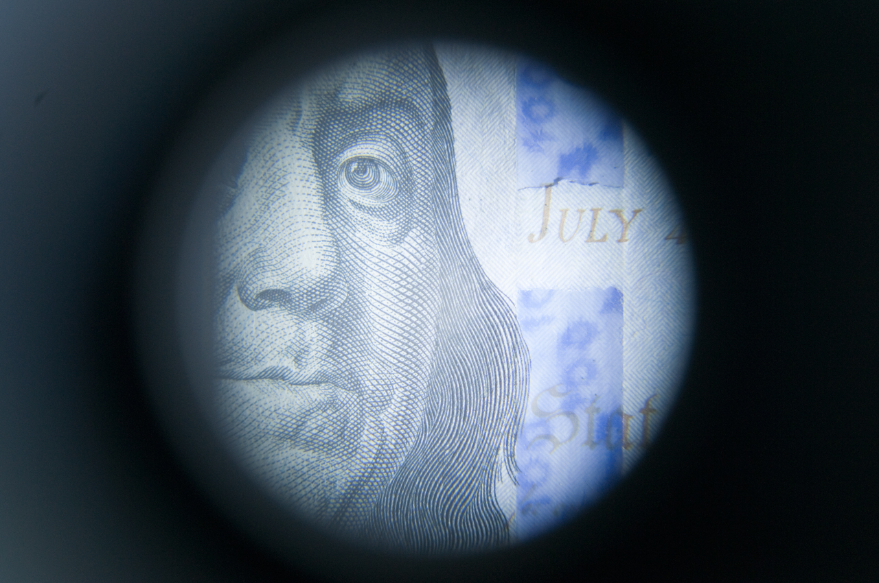
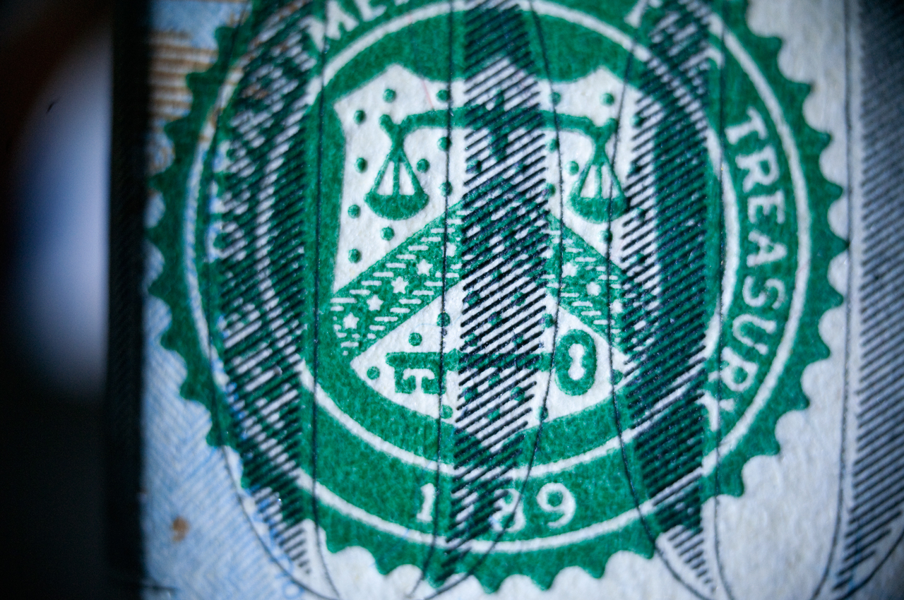
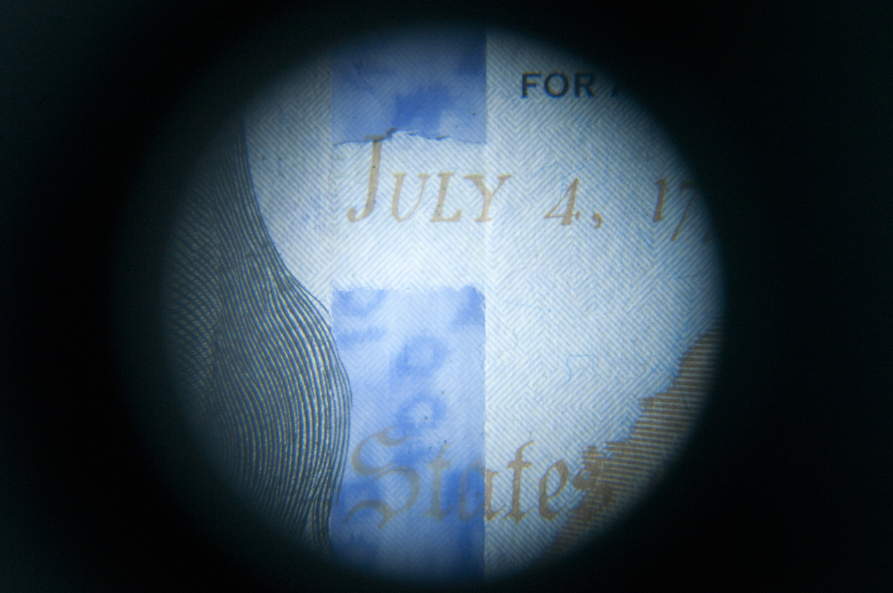
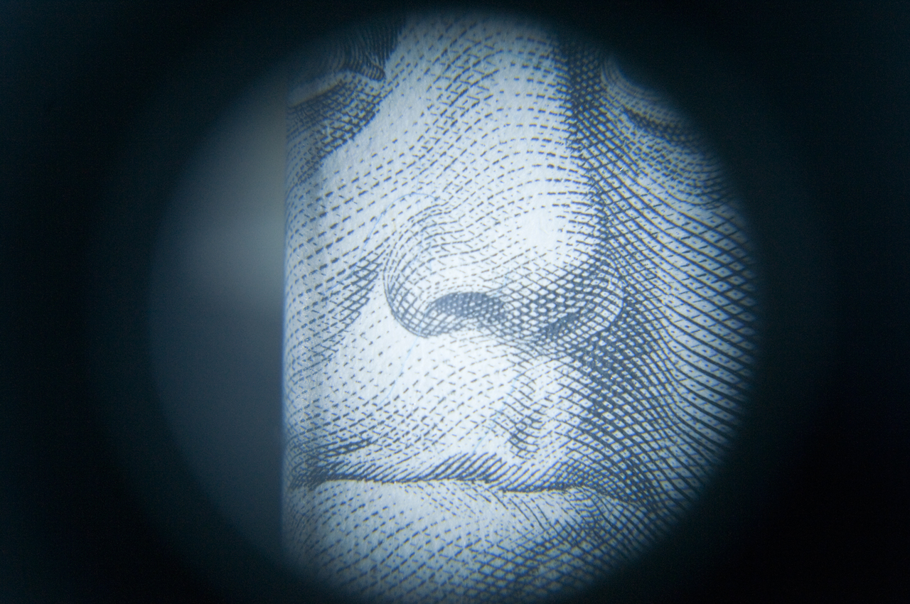
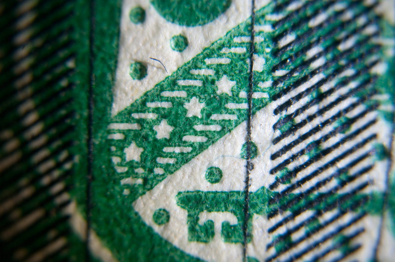
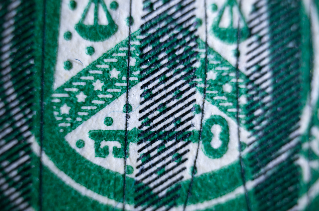
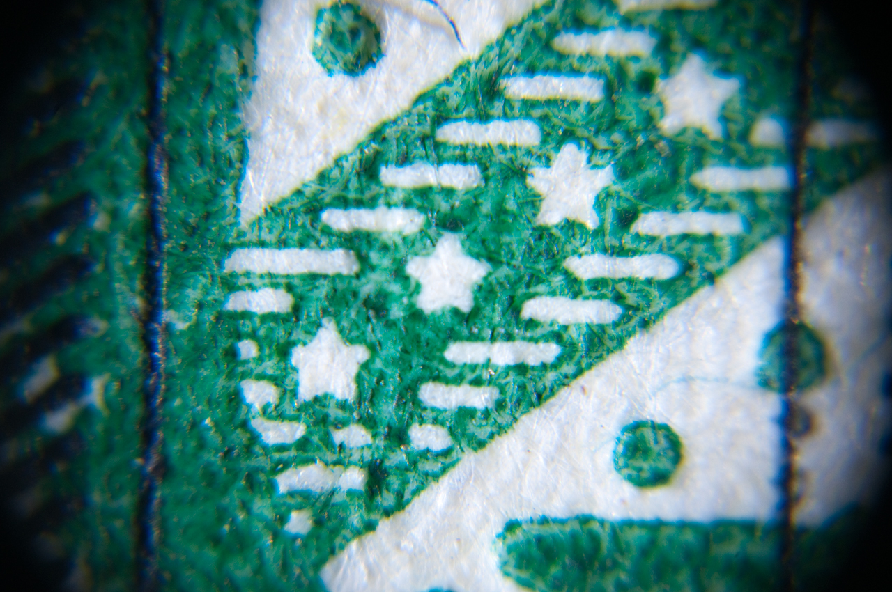
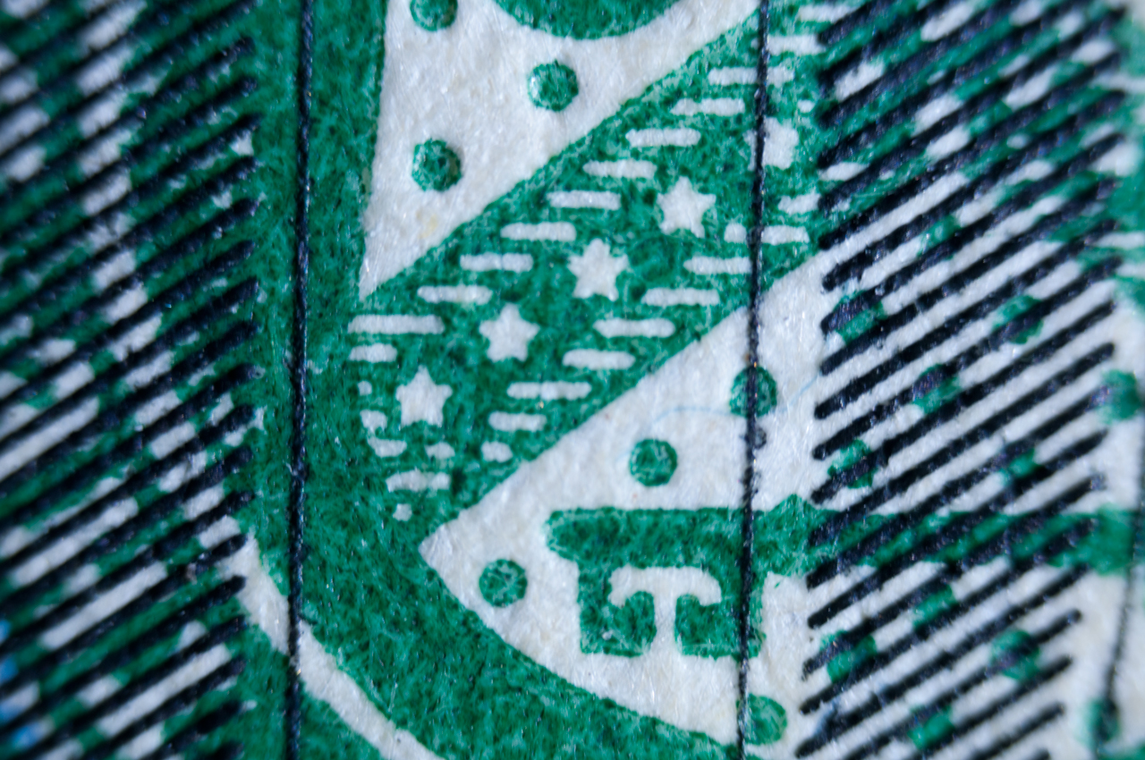
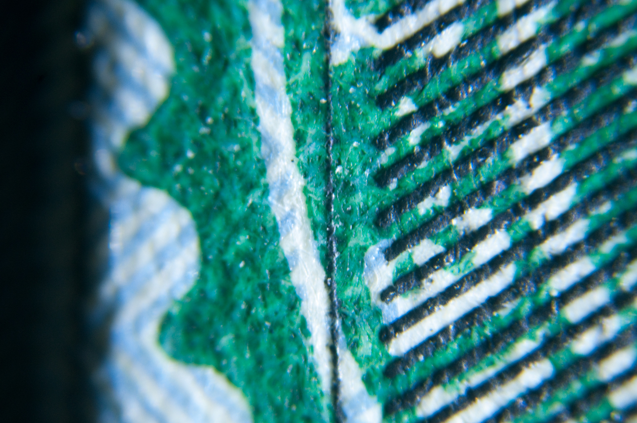
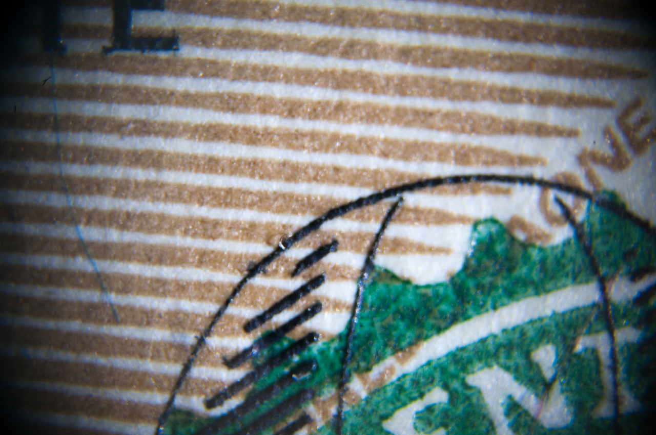
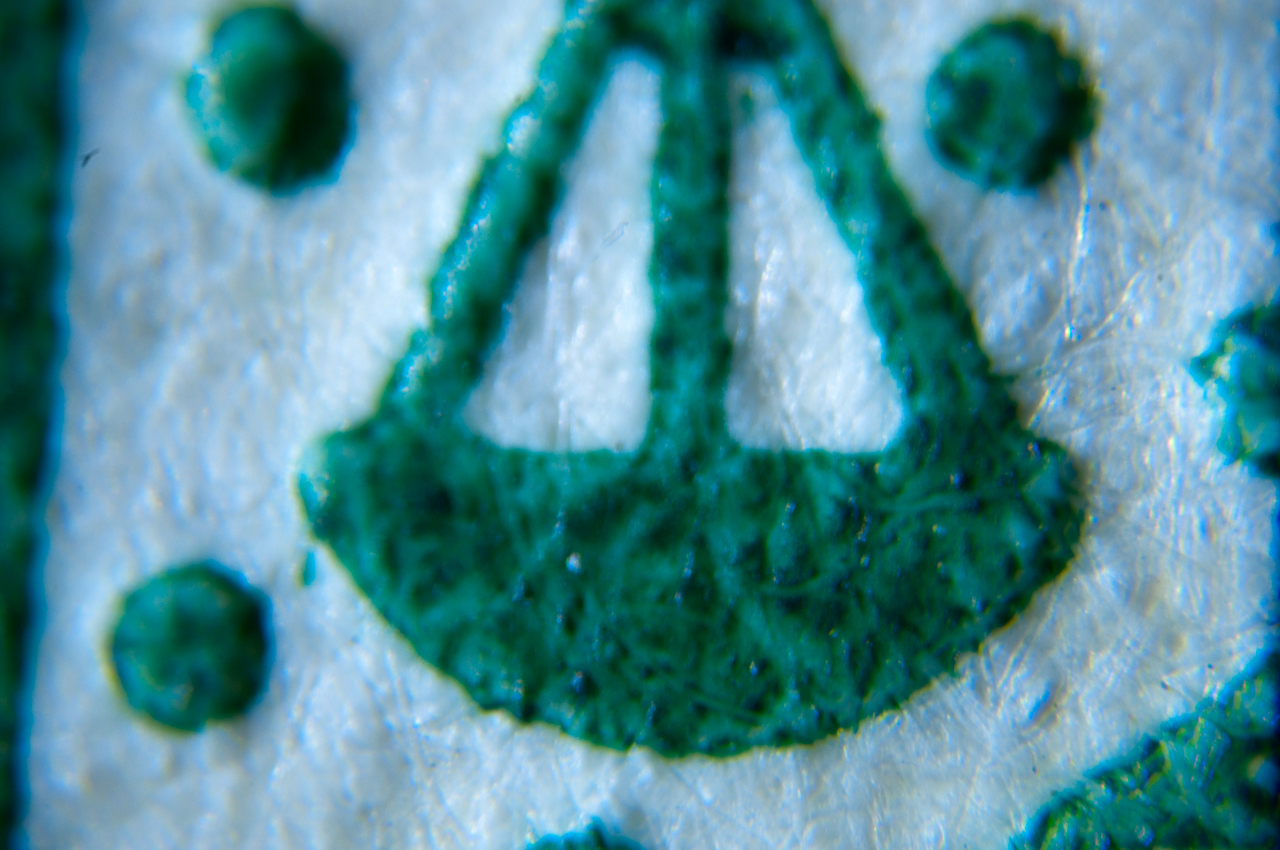
Photo of front of U.S> $100 bill (new issue 2014).
Flashlight at about 45 degrees for light.
Nikon D300s Manual mode adjust shutter for camera recommended
exposure (ISO at 200).
Lens code: [2 letters maker)(power followed by X or XI if
infinity)(cg if for cover glass) (nnn extension tube length in
mm).
Taken with camera sitting on table top and bill held onto a 123
block with rubber bands.
Focus by using live view and a couple of presses of the magnify
button, but it could be better if the camera was better
anchored.
Notice extension tubes only reach to 98mm (+3mm camera &
objective adapters for 101 mm total) not 160mm, need more
extension tubes or a custom tube.
Under each photograph is the exposure time (ISO: 200).
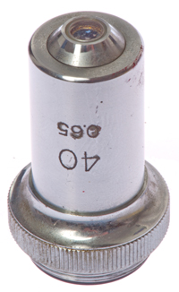
It's difficult to focus the 40X objective by moving the camera,
need a stand with slow motion control.
Note: In the eBay ads for these adapters they typically
show a camera and no extension tubes then a microscope
objective. The images you get are the first row in
the table below and are not what the objectives were designed to
do.
Note the Flange to Focal distance (Wiki) for
the Nikon-F is 46.50 mm.
This is the distance that needs to be added to the extension
tube length to get the working focal length.
If the real image is formed 10mm down the main tube so that it
is inside the eyepiece then the real image is formed about 150mm
from the objective flange.
The last row in the table is for a working focal length of
147.5mm which is pretty close to the desired 150mm.
Note1: The Labophot microscope focusing stage will not go up to a
parafocal distance of 31mm (RMS) and that may be why some of the
objectives do no focus.
Note 2: With the objective mounted close to the camera the image
circle is smaller than when it's out at 160mm so you can get some
idea of the image circle.
Tube Lens
A tube lens is used with the Nikon CFI (Chrome Free Infinity)
optics system. One way to use the CFI objectives with a
camera is by placing them in front of a telephoto lens using a
filter thread to 25-0.75mm adapter. But I'd like to
incorporate a true tube lens into an optical adapter
system. Since a classical RMS microscope tube takes
eyepieces that are 23.2mm in diameter it's a little too small
for tube lens optics, so I'm using T-mount parts which have an
ID greater than 30mm. C-mount works OK for RMS tube size
optics hence most of the camera
to microscope adapters use C-mount parts.
Note: The T-mount threads (should be M42-0.75) on the
Edmund Optics parts do not mate with theM42 on any of the Made
in China parts.
The problem is with the M42 Made in China parts is that they
are using M42-1.0 lens mount threads, not the M42-0.75 threads
that are specified for T-mounts.
Note: If a thread is specified just a M2 thread then
it's the M42 Lens thread (Wiki)
which is M42-1.0.
If a thread is specified as T-mount (Wiki) then
it's the M42-0.75 thread.
It's easy to tell the difference using a metric thread pitch gauge.
Edmund Optics sells a "Nikon 200mm tube lens" that's mounted in
a 38mm threaded housing p/n: 58-520,
but it's $250. It's possible the actual lens is p/n: 45-415?
The 45-415
lens is used as the objective lens in the Edmund Optics Cage
System Autocollimator
Fig TL1 T-Mount (42mm-0.77) Tube Lens
Housing & Lenses
Anchor Optics (the discount part of Edmund Optics) has
some
lenses that are suitable for use as a tube lens.
 |
Fig TL2 100mm Ext & stack of two sets
of 3
each about 100mm tall.
Edmund
Optics 52-296 100mm T2-mount extension on left
Made in China M42 (-1.0) extension tubes on right
Note: T-mount on left, M42 on right.
|
Fig TL3
T-mount Lens holder
with Tube lens installed
Edmund
Optics 57-977
In this photo the lens is
1mm from the front of
the top mating surface.
It's also about 10.5mm
from the bottom mating
surface.
|
Fig TL4 Nikon CF Plan 10X ∞ objective & 118mm FL tube
lens
& 100mm T2 ext tube.
It's difficult to get the focus set and
make the image sensor be parallel with subject.
But this is the best image so far! (June 2014)
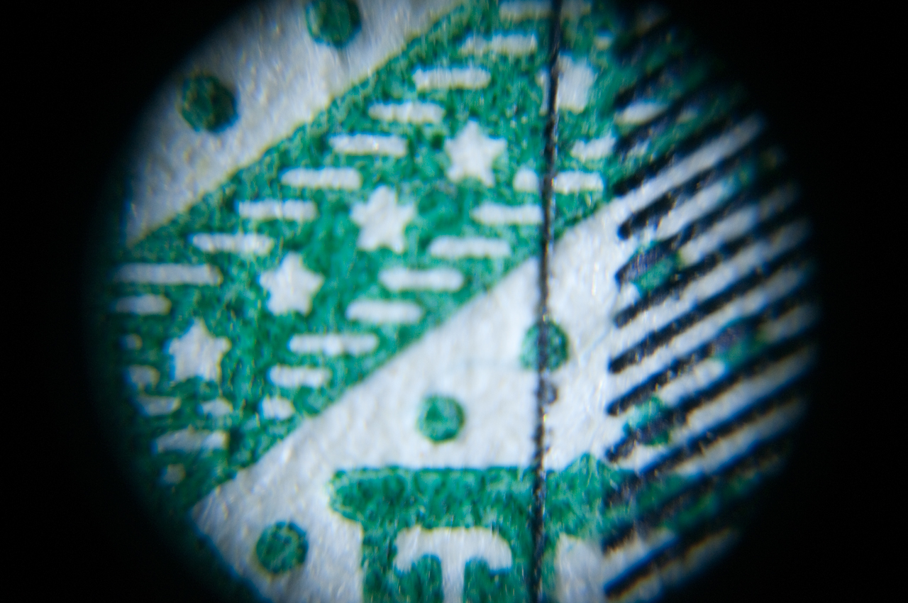
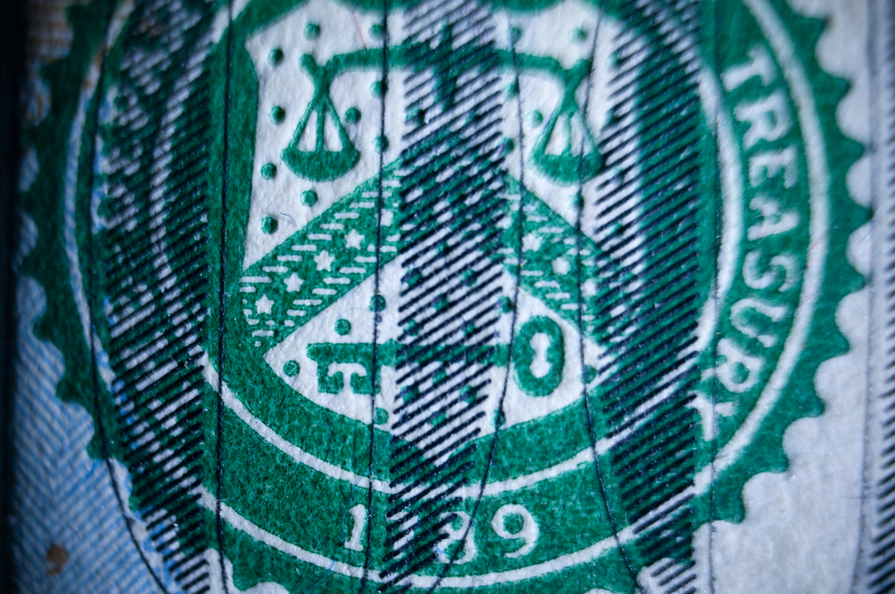
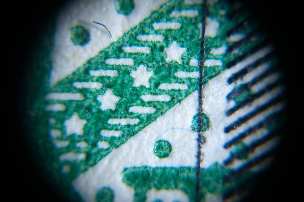
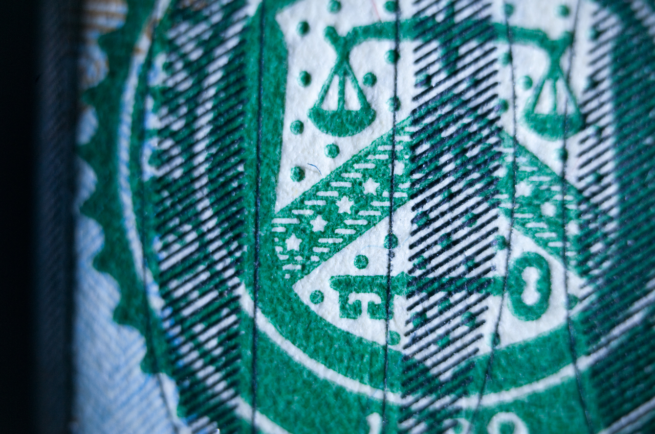
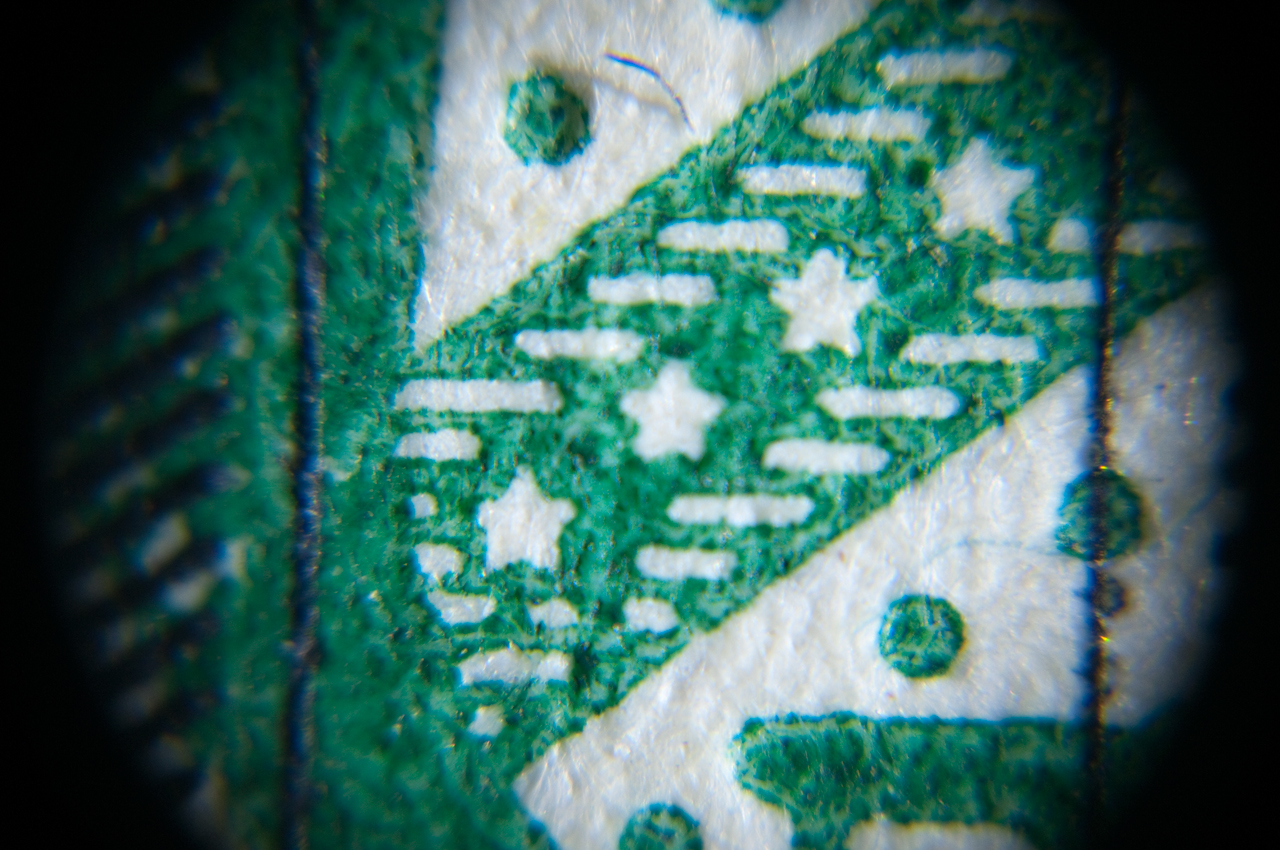
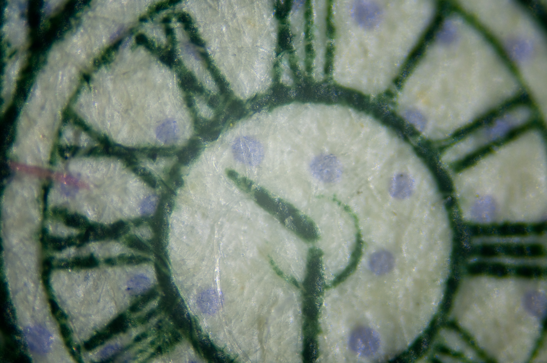
Notice the blue dots (new $100 bill back side)

|
Fig TL5 D60 for illustration, actual photos
taken with D300s.
Note front 42mm to RMS adapter does not match T-mount
threads
on lens mount so there is a gap.
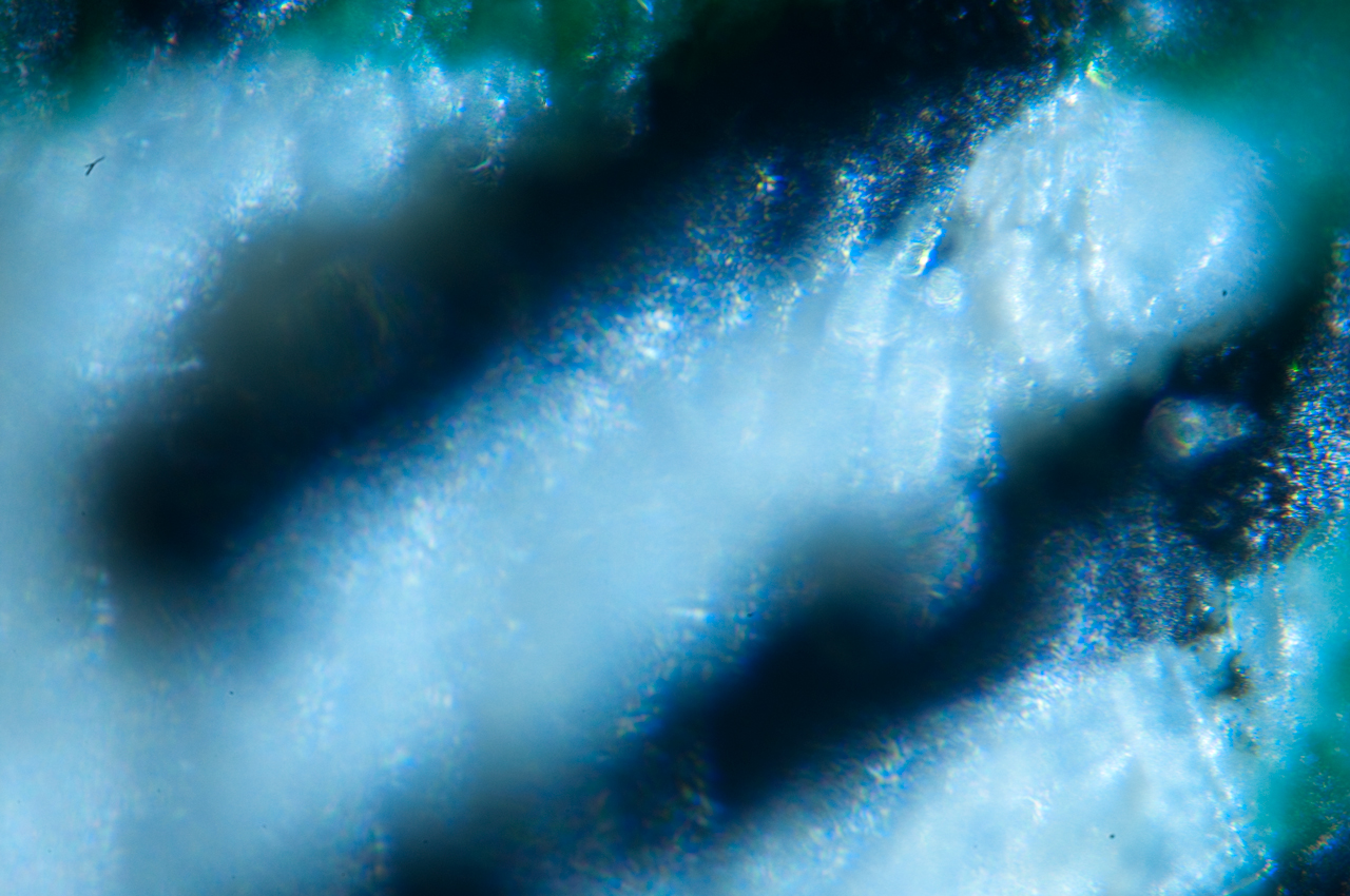
With this setup a photo of the mountains is all blurry,
i.e. this setup
has the tube lens out of focus.
|
Fig TL17 Camera with tube lens with
objective adapter
and objective removed. It's now a 200mm telephoto
lens.
Focus is set to infinity and locked with setscrew.
Photo taken with Nikon D60.
Nikon to t-mount adapter
100mm T-mount extension tube
Focusing T-mount adapter
T-mount lens holder for 30mm dia lens holding tube lens.
|
Fig TL6 with bellows to allow focusing.
Fig TL5 shows the tube lens too far from the camera to be
focused.

|
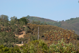
Fig TL7 image of mountains with setup shown
in Fig TL6.

|
TL 20 same as T-17 above but with ISO
microscope adapter.
ISO is a 38mm (1-1/2") camera interface for microscopes.
|
Fig TL8 Nikon CF Plan 10X ∞ objective & 118mm FL tube
lens & bellows
NOTE: The bellows rails are in the way of where the
subject wants to be.
|
The objective to camera flange distance can
be measured directly
when the bellows is removed from the camera and is
89.75mm.
When the Nikon-F flange to focus distance of 46.50 mm
is added
to that it comes out 136.25mm, but the nominal FL of the
tube lens
is 118mm so there's a discrepancy of 18.25mm. That's
because the
tube lens sits back from the objective flange pretty much
by that
amount.
If instead of measuring camera flange to back of objective
the
distance from the camera flange to the front of the
T-mount adapter
is measured the result is 65.25mm. Adding 1mm for
the lens shelf
and 46.50mm for the camera focal dist gives 112.75 which
is closer.
Maybe the center of the lens is that far from the edge?
|
|
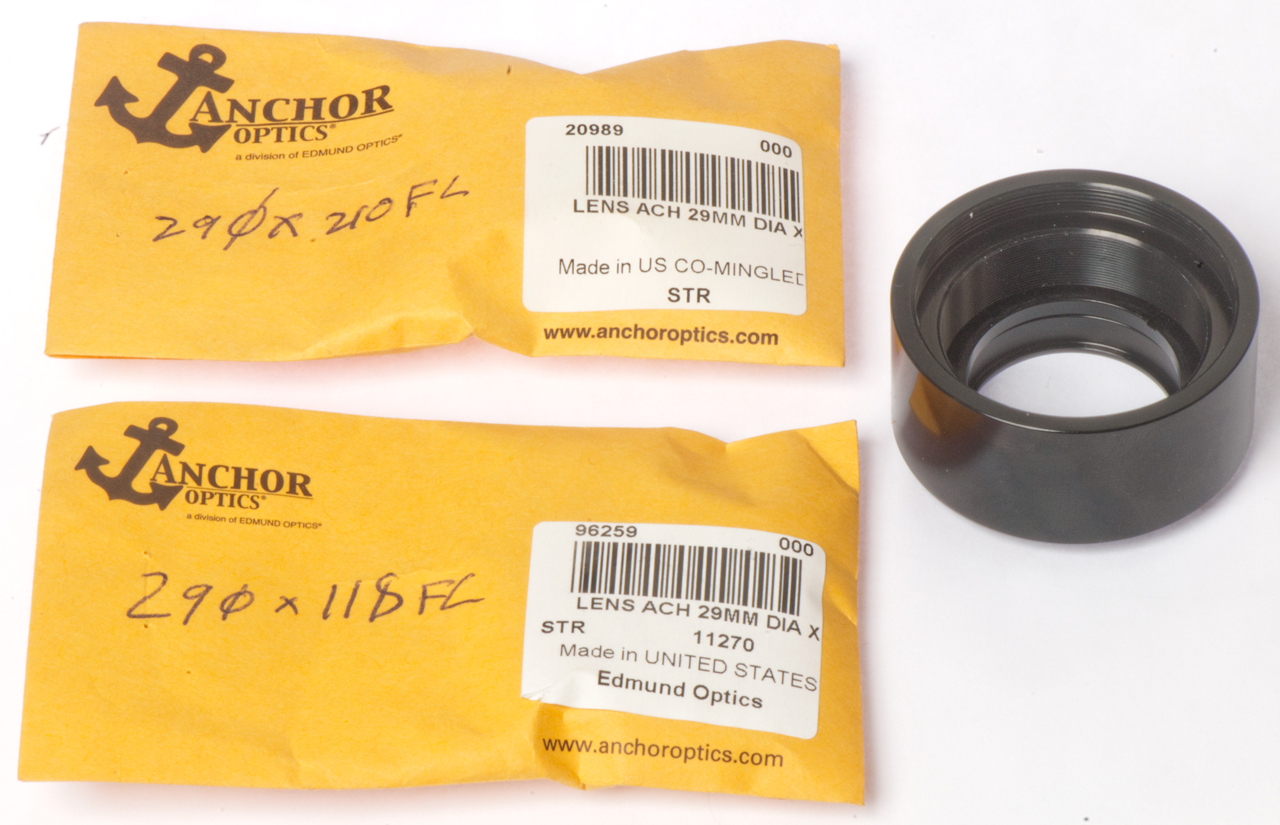
On order is Edmund Optics p/n: 45-415
MgF2 Coating, Achromatic Doublet Lens
200mm FL x 30mm dia lens to fit
existing 57-977 T-mount thick lens holder
10mm T-mount Ext tube p/n: 52-294
40 to 65mm adj T-mount ext tube p/n: 58-755
|
|
|
Fig TL9
New 45-415 Tube lens mounted in 57-977 thick lens T-mount.
100mm + 40-65mm T-mount extension tubes.
After removing T to RMS adapter and objective,
focused at infinity (local mountains).
17 July 2014 Note on Edmund 40 to 65mm adj T-mount ext
tube p/n: 58-755:
There is a 1.5mm pinch screw to lock adjustment.
There is an internal 32mm ID position for a lens.
The retaining ring has an ID of 29mm, so could be used for
the 30mm tube lens.
|
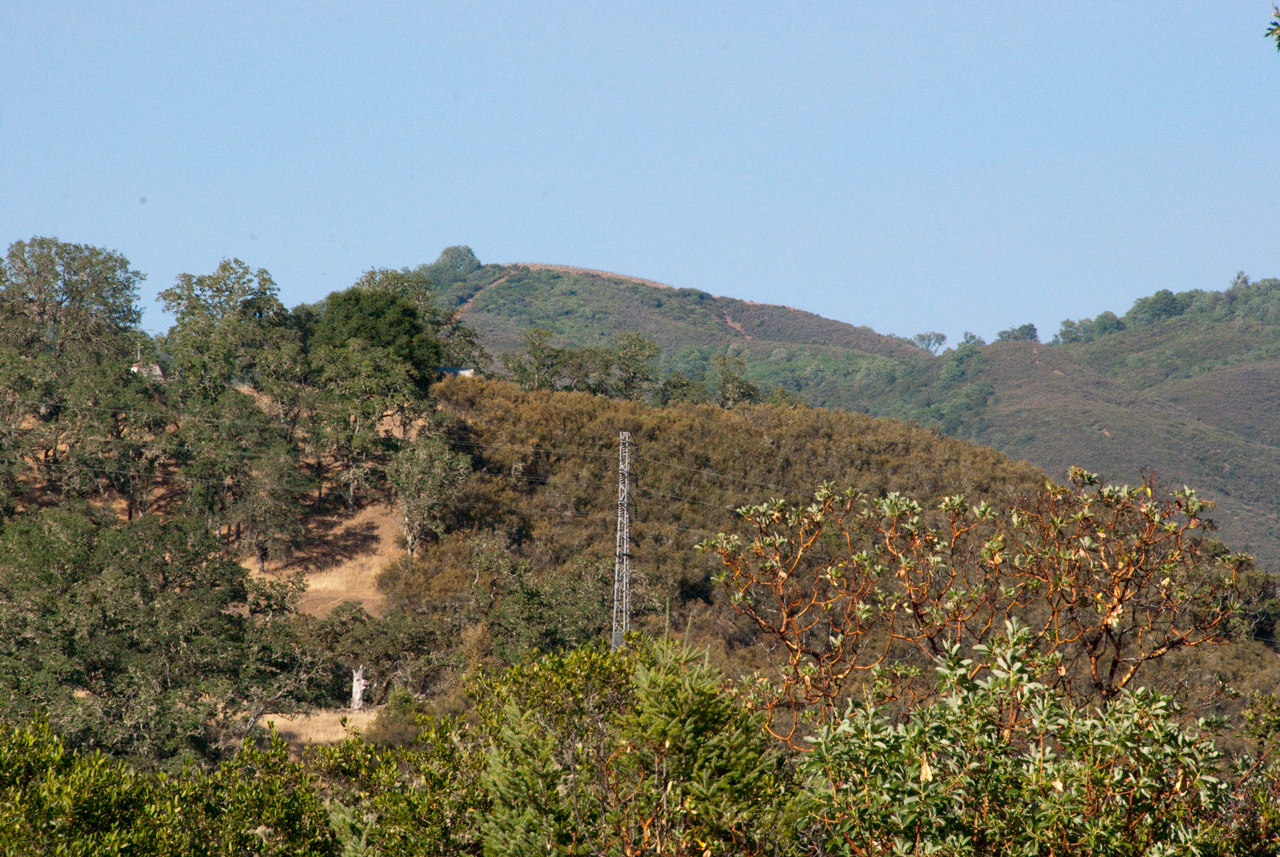
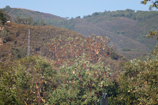
Fig TL10
Local mountains
200mm tube lens 1/640 sec, ISO 200
|
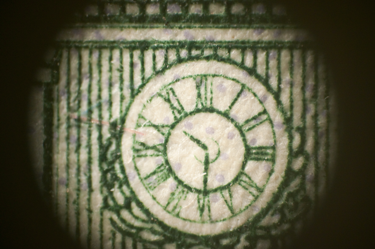
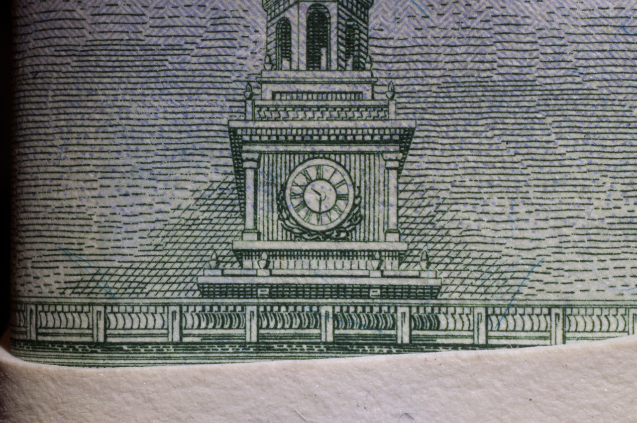
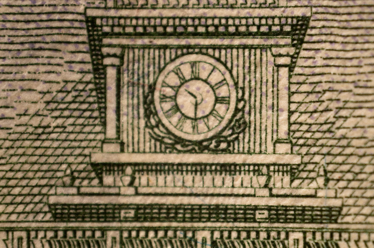
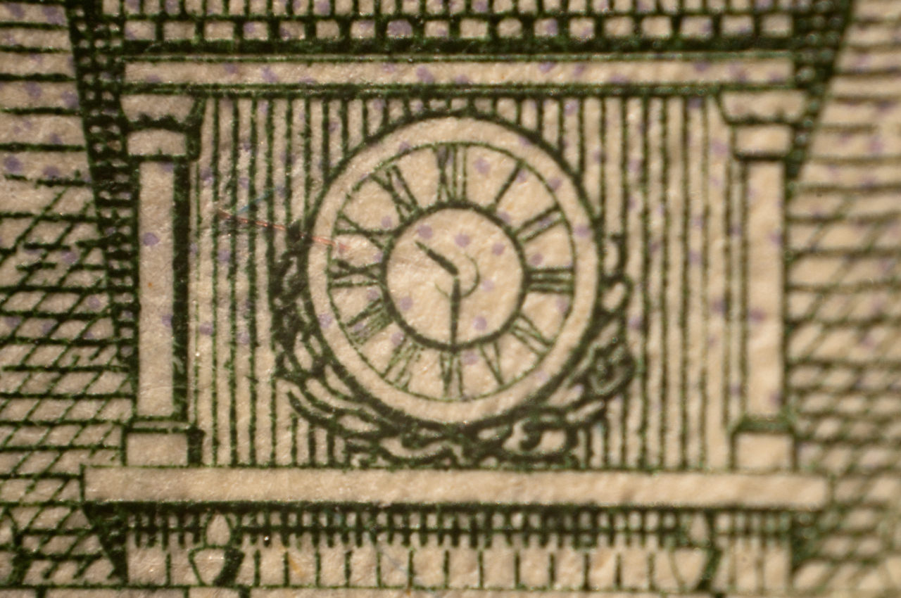
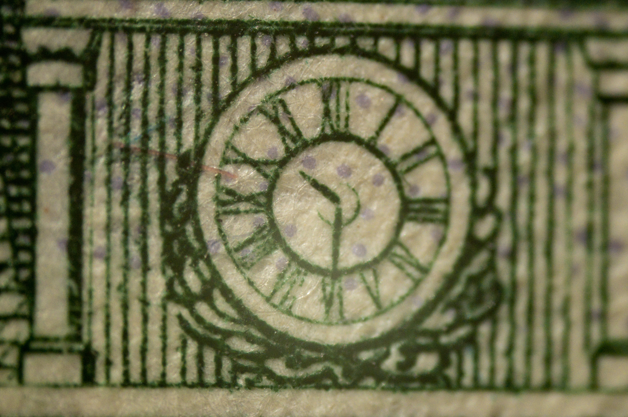
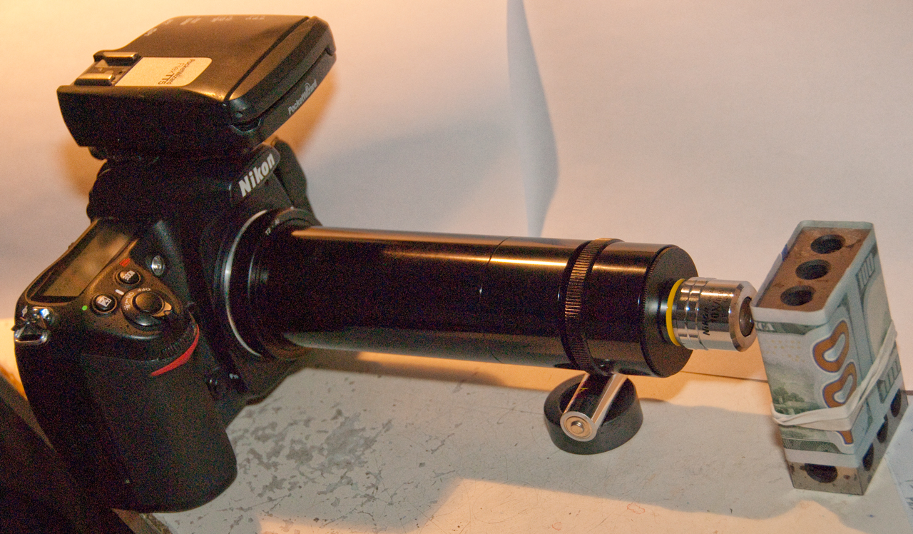
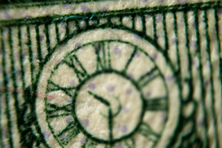
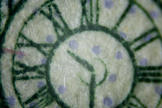
Fig TL11
100 dollar bill clock
Taken with setup in Fig TL9
Note magnification is higher with 200mm tube lens in this
photo
compared with Fig TL8 taken with 119mm tube lens.
|
TL 12 Actual camera and test subject for
Fig TL11

|
Fig TL13 taken with Nikon D60 + 18-200mm DX
ED lens set to 1/640 sec, same ISO and shutter speed as in
Fig TL10.
Data shows f/22.
|
|
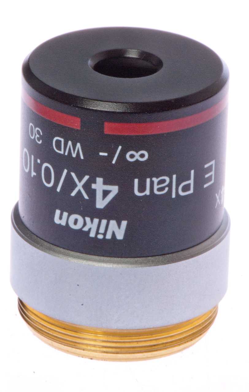
TL 14 D300s, 80-210mm Lens, 52mm-25mm, E
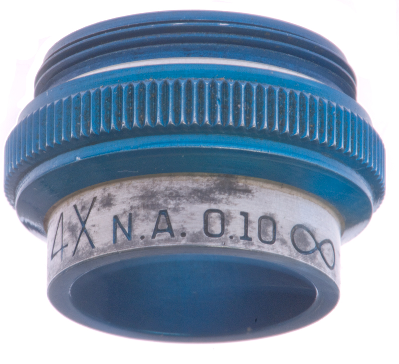
Plan 4x/0.10 ∞ / - objective Note:
Manual focus mode and set to ∞.
52mm
to 25mm adapter from cnscope
|
|
|
A tube lens is supposed to be needed when using infinity type
objectives. So I'm going to do some experiments making
one. The tube lens is to both allow more room in a
microscope for additional optical elements, like filter cubes,
and to provide some image compensation to improve image
quality. To go along with this some objective lenses use a
25mm thread instead of the smaller RMS thread to allow higher
N.A. lenses that can be made using the RMS thread. The
Nikon CFInn series of objectives in an example.
There are a number of different "mounts" that might be used.
C-mount (Wiki)
is intended for movie and video camera lenses (it has a 1"
thread size so the hole is smaller than that), but it might
cause vignetting of the image on a full size 35mm camera.
The S-Mount (Wiki)
is for very small board cameras and has a 12mm thread so is much
too small.
T-mount vs. C-mount
Most of the parts I'm using for this tube lens experimentation
are also available as C-mount which works with TV/CCTV/Video
camera lenses and is smaller in diameter and lower is
cost. I've chosen to work with T-mount because the lens
diameters are larger thus have faster f/numbers and maybe will
be working more near the center than the edges both of which
should lead to higher quality images. The max lens
diameter that will fit in a stock C-mount lens holder is 25.4mm
(1"), so the 45-415 will not work in a C-mount system.
This will be done by mounting a lens in a T-mount (Wiki, M42-0.75
threads) tube so that the distance between the objective
and tube lens and between the tube lens and camera imaging chip
can be adjusted.
From the Edmund Optics catalog B141C page 315:
D1(mm) = 2 * F1 * N.A.
[Equation 1]
L(mm) = [(D2 - D1) * F2] /
D [Equation 2]
Where:
L = dist between objective and tube lenses
D = Field diameter
D1 = Objective Exit Pupil diameter (mm)
D2 = Tube Lens Entrance Pupil diameter (mm)
F1 = Objective Focal Length (mm) is a function of the
tube lens focal length, see Eq 1 above.
F2 = Tube Lens Focal Length (mm)
N.A. = Objective Numerical Aperture
So, when using the Spencer, Cat. 1076, 10X/0.25, Infinity lens
with two different tube lenses here are some calculations.
Entrance pupil = 5.23 mm by measurement, not optically
calculated
D1 =Exit pupil = 9.1mm by measurement, not optically
calculated
The Nikon D300s (Nikon
data)has an imaging chip size of: 23.6 x 15.8 mm so D is
about 15mm.
There are 4288 x 2848pixels on the chip.
Each pixel is 0.005504 x 0.005548mm or 5.5 x 5.5 um
The two tube lenses are shown in the table below. Note
these are low cost lenses, there are very nice lenses available
with good coatings for much more money.
Edmund Optics Lens p/n:
|
96259
|
20989
|
45-415
|
| F2 Focal Length Tube Lens
(mm) |
118
|
210
|
200
|
Tube Lens O.D. (mm)
|
29
|
29
|
30
|
D2 = Tube Lens Entrance
Pupil (E.O. p/n
57-977) (mm)
|
28
|
28
|
|
| F1 = Tube Lens Focal Len / 10x |
11.8
|
21.0
|
|
| D1 = 2 * F1 * 0.25. |
5.9
|
10.5*
|
|
L = [28 - 9.1) * F2] / 15
|
149
|
264
|
|
Note * The calculated exit pupil is larger than the physical
hole in the objective, this may be a problem?
Nikon
CFI60 Optical System - All of
the CFI60 objectives use a 25mm thread rather than the RMS
thread.
Basic
Principles of Infinity Optical Systems - includes a flash
calculator with Tube Lens to image distance (160 to 200mm) and
Objective Focal Length 2 - 40mm (Power 80 to 4) as variables.
Infinity Head (built-in tube lens)
This infinity head came from an unknown microscope. It
has a 38mm dovetail, smaller than the Nikon CF head 48mm
dovetail.
After installing Nikon CFW10 eyepieces and taking the head
outside and pointing it to the distant mountain ridge it was
almost in focus.
Extending the eyepiece focus almost all the way out brought the
ridge into focus. Thus confirming that this is an infinity
head.
The entrance pupil diameter on this head is 9mm and on the Nikon
CF head it's 14mm.
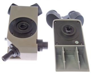
Infinity Head Fig 1
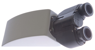
|
Infinity Head vs. Nikon CF Head
|
Epi Illumination
It's possible to illuminate the subject using the
objective lens as a condenser (it inherently has the same N.A.
as a condenser as it has an an objective). This can be
done using a beam splitter between the objective and tube
lens. Light is fed from the side of the beam splitter,
reflects to the objective and the light from the objective goes
to the camera. Possible parts are:
Edmund Optics T-mount for beam splitter p/n: 63-981
40mm 50R/50T cube beam splitter p/n: 32-506
In addition there needs to be a collimated light source to drive
the third input port so the cost is going to be over $1,000.
Infinity
Microscope Optics Patents
725839
Apparatus for Facilitating the Sighting of Distant Objects from
Submarine Boats, Barbettes, &c. Howard
Grubb, Dec 13, 1901, 359/405
Parallel light rays travel down the tube. Grubb
mentiones looking up the tube and you will see an image at
infinity. But the telescope objective for the bottom part
much be above the eyepiece at an optical distance of it's focal
length, i.e. it's focused at infinity.
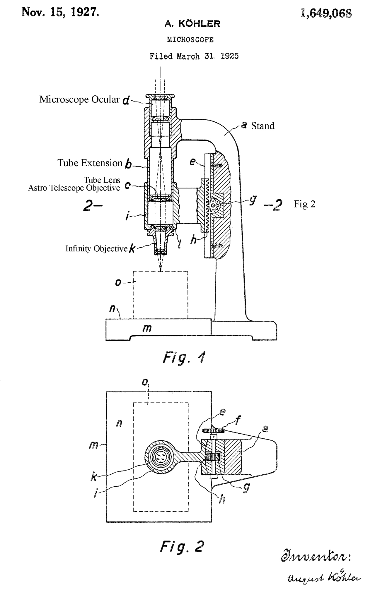
1649068
Microscope,
August
Köhler, Zeiss
Carl Fa, Apr 16, 1924, 359/379 -
The stand (a) holds in a fixed
position the ocular (d) and substage (o).
Only the infinity objective (k) moves.
Instead of using the term "tube lens" the patent
calls it an astronomical telescope objective (c).
So the combination of the ocular (d) and tube lens
(c) makes an astronomical telescope which is
focused at infinity. This means that if you
remove the objective lens and it's mounting plate
and have just the camera and tube lens
installed you should be able to take photos
of subjects that are at infinity. Removing
the stereo head from an infinity tube length
microscope allows using the head like a pair of
binoculars.
My first try at an infinity tube system for system
was very fuzzy. The 100mm T-mount extension
was too long so not focused at infinity.
But after reading this patent it's clear that the
head must act as an astronomical telescope, that's
so say a telescope focused at infinity.
|
 |
|
3132200
Microscope
optical system, American
Optical Corp, Jun 5, 1961, 359/379,
359/763
- aimed at having tube and stage fixed to frame an only moving
nose for focus.
Fig CoM4 Nikon D60
(for illustration only)
With Nikon-T adapter and T-23mm adapter
On Unitron microscope.
|
Fig CoM1
1. Nikon to T-mount adapter
2. T-Mount to 23mm microscope eyepiece tube adapter w/2
lenses
3. 23mm eyepiece tube to 30mm eyepiece tube adapter

|
Fig CoM2
same as Fig CoM1 except showing bottom of T-Mount to 23mm
and second lens.
 |
Fig CoM5
|
Fig CoM3 adapters mounted on camera.
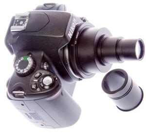 |
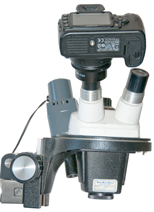
Fig CoM6 on Mitutoyo Toolmakers
Measuring Microscope
 |
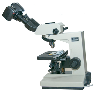
Fig CoM7 Nikon Labophot
Microscope
See Example image
No. 1
|
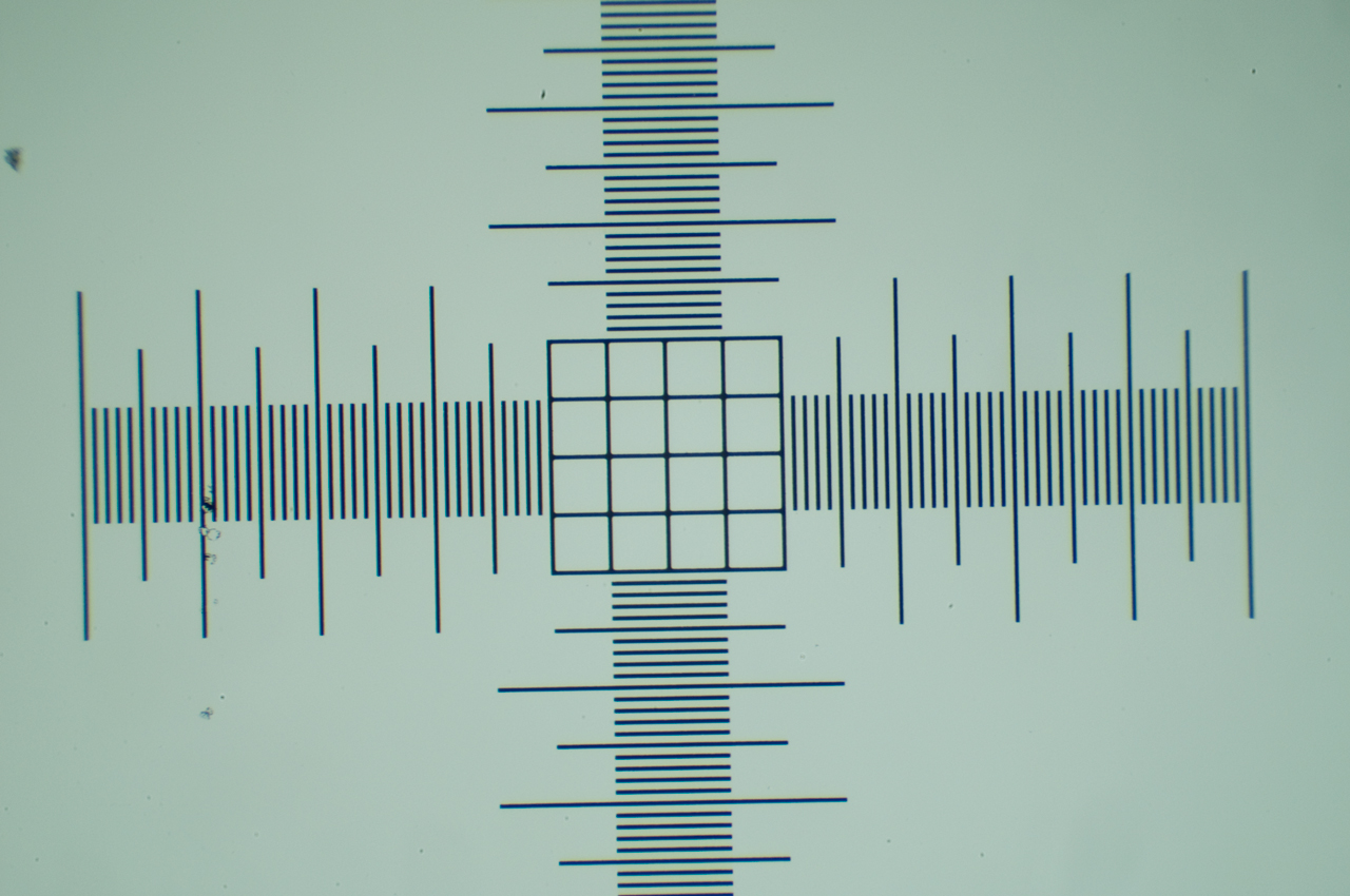
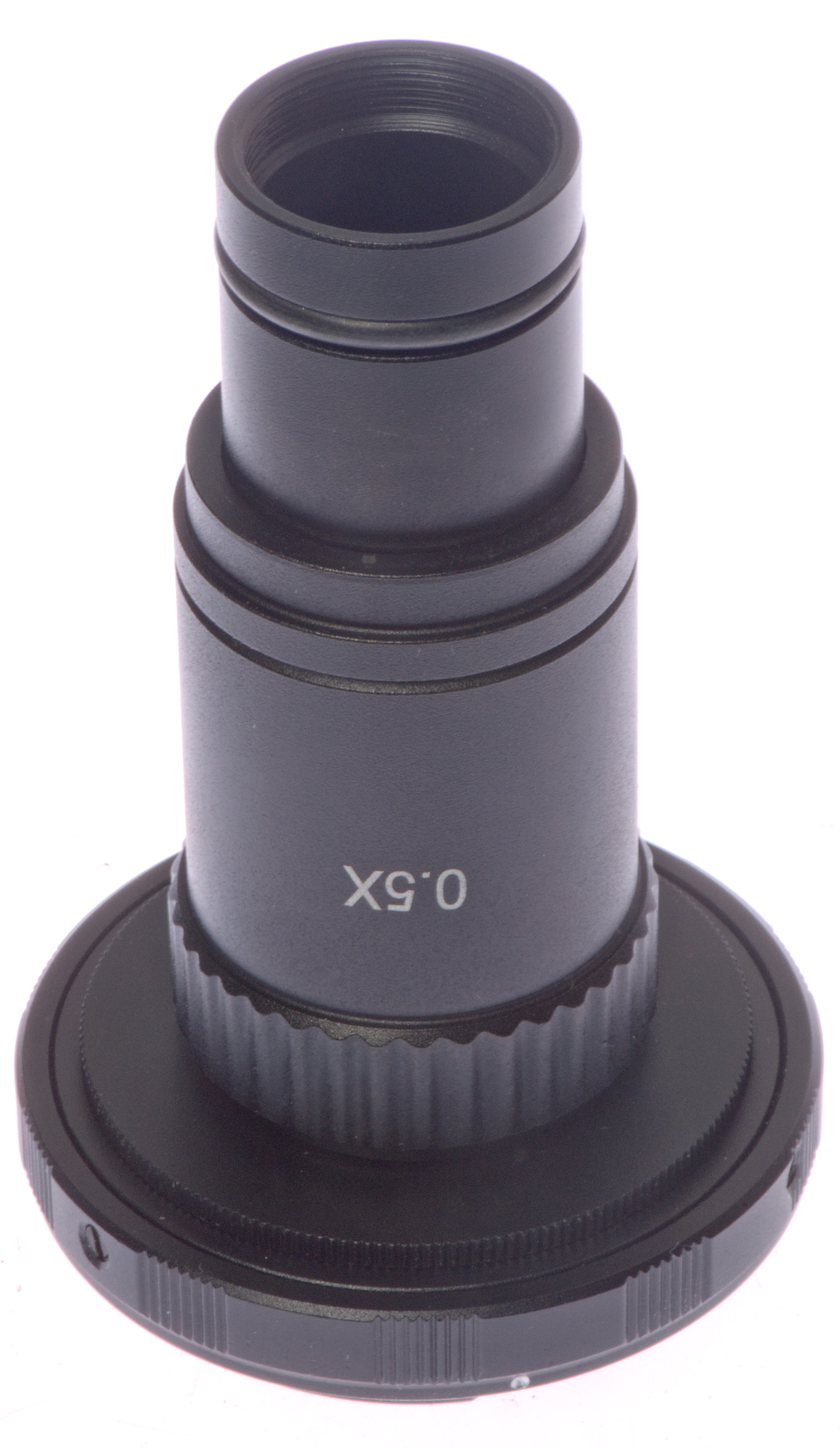
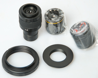
Camera Adapter sold as 0.5x
1230 pixels * 5.5 um/pixel = 6765 um = 6.765mm
6.765 / 5x objective = 1.353 power camera adapter
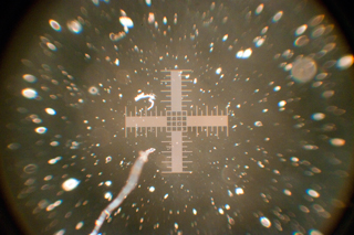 |
In Labophot using light from below 10x E
Plan objective
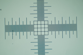
From left to right in Photoshop = 3694 pixels canvas is
4288 pixels
3694 * 5.5 um = 20.3mm
3694/4288 * 23.6mm = 20.3mm
|
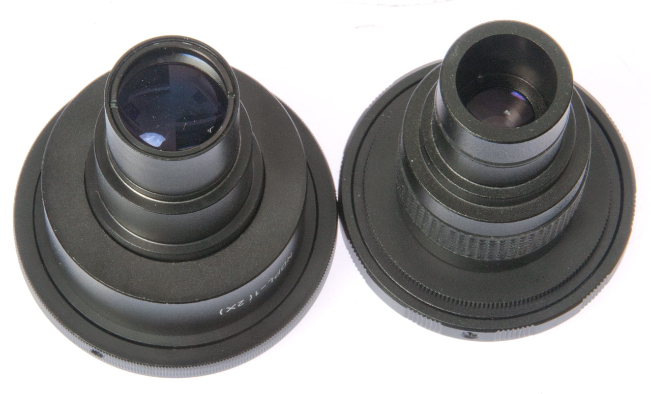
Optexcom.com Camera Adapter
From eBay seller newhoper.
Marked as a 1x relay lens that comes with a Nikon to T-mount
adapter, and a T-mount to C-mount adapter and the actual optics
in a housing with a 23mm nose and C-mount output, along with a
couple of 23mm to other size eyepiece tube adapters (29.9mm
yellow band, 30.5mm).
The magnification of both this (Opterexcom) and the above
Imaging Solutions relay lenses is very close to the same
magnification, the Optexcom shows vignetting that is not in the
Imaging Solutions relay lens.
|
3719 pixels * 5.5um = 21.54mm
|
Comparing Image Solutions and Optexcom
Camera Adapters
Although the Image Solutions relay lens is marked 2x and the
Optexcom is marked 1x, they have essentially the same
magnification.
By measuring Vernier calipers set to 0.5mm (1.0mm is too wide),
see photo below the true magnification can be shown to be 2X for
both relay lenses.
So the Optexcom lens is mismarked at 1X, it's really 2x.
The diameter of the lenses used in the Imaging Solutions camera
adapter are considerably larger.
The big advantage of this method is being able to utilize modern
lighting methods and the big disadvantage is the high cost of a
microscope that does not have an eyepiece that moves as the scope
is focused. The accessories needed for each of the lighting
methods and special objectives for each lighting method also add
expense.
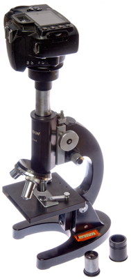
Note that it's very difficult to mount a digital SLR camera on top
of an older microscope using an eyepiece adapter. The
problem is that old microscopes move the tube up and down to focus
and the camera weight drives the tube down and can break the slide
and/or damage the objective. Modern microscopes mount the
camera to a fixed part of the microscope frame so this is not a
problem.
There are two optical approaches to doing this.
Afocal (Wiki)
where the camera is focused at infinity and takes a photo through
the eyepiece lens. Nikon made an adapter (Model 2?) in
two parts where the eyepiece was removed, the lower part was
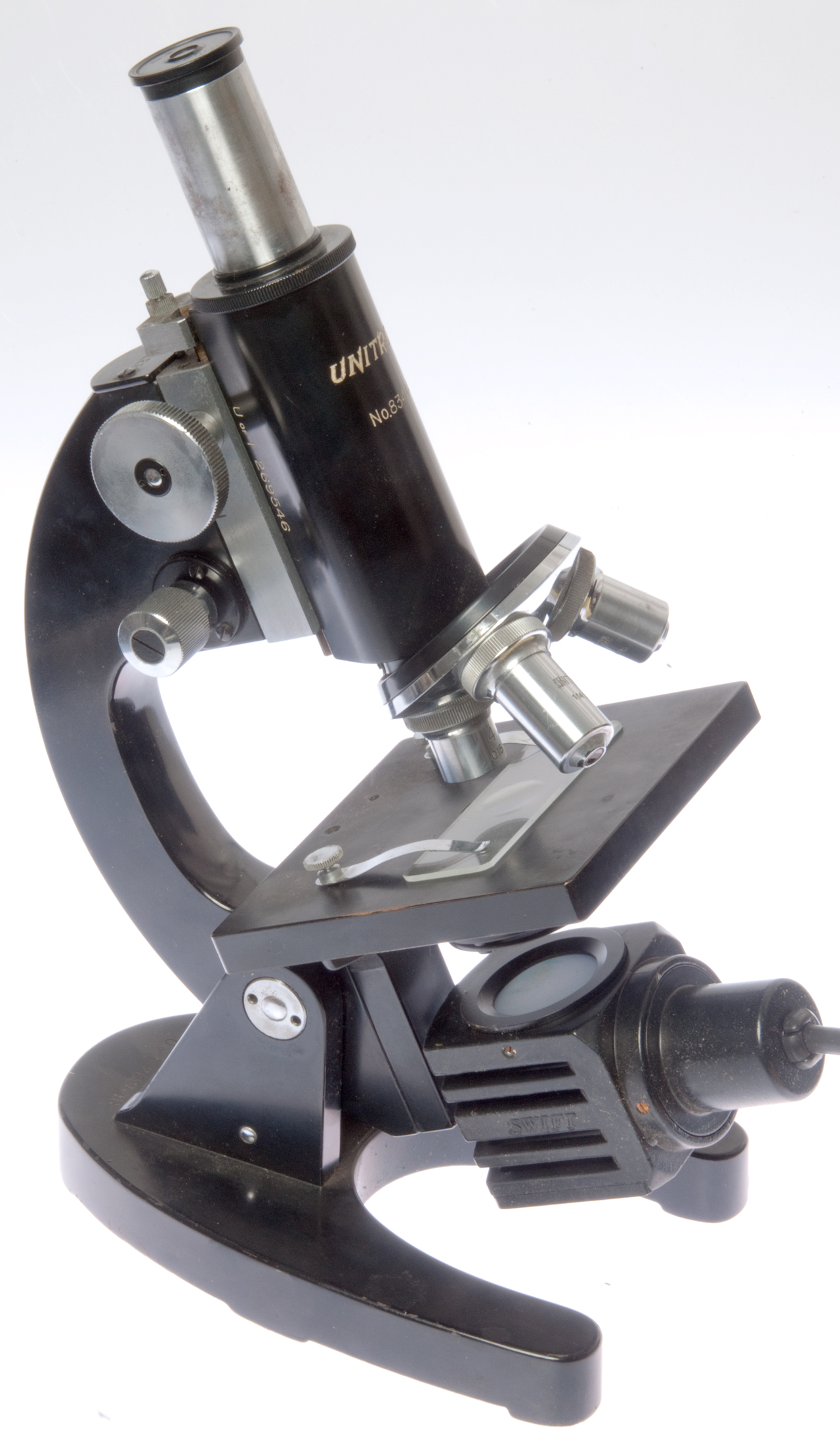
clamped to the microscope barrel, like the Unitron below, the
eyepiece was reinstalled, then the upper part was installed with
the camera.
In eyepiece replacement the microscope eyepiece is removed and
replaced by the camera with an adapter (typically 23mm). I
think these need a relay lens since the focal plane for a real
image is down inside the microscope tube and that needs to be
relayed up to the imaging chip in the camera.